
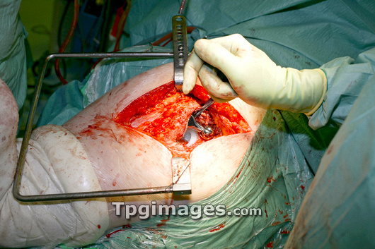
MODEL RELEASED. Hip replacement surgery. Image 7 of 9. Surgeon manipulating the head (metal ball) of a prosthetic hip joint into its prosthetic socket (white) during hip replacement surgery. The head is attached to a stem inserted in the femur (thigh bone). The socket sits in the pelvis. The prosthetic joint will articulate in exactly the same way as a normal hip joint. Joints may need to be replaced after injury, disease or deformity. This hip is being replaced because of congenital hip dysplasia, a condition where the hip joint does not fit together properly, causing it to dislocate frequently and to become worn. For a sequence showing the operation see M551/418- M551/426.
| px | px | dpi | = | cm | x | cm | = | MB |
Details
Creative#:
TOP03217465
Source:
達志影像
Authorization Type:
RM
Release Information:
須由TPG 完整授權
Model Release:
Y
Property Release:
N/A
Right to Privacy:
No
Same folder images:
 Loading