
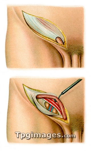
Inguinal hernia surgery. Image 1 of 2. Historical artwork of the first two stages in the Bassani technique to treat inguinal hernias, where the intestines protrude into the inguinal canal. In the first stage (top) the skin and fat layers (yellow) have been cut open to expose the membranes (white) covering the surface muscles. In the second stage (bottom), the muscle and connective tissue layers have been cut open, and the spermatic cord (red) retracted with a hook. This has revealed the herniated peritoneum (abdominal wall), with the intestines (brown, with red and blue blood vessels) protruding outwards. Artwork from Atlas and Epitome of Operative Surgery (1898, Otto Zuckerkandl). For a sequence of images showing the four stages of the operation, see images N700/098 and N700/099.
| px | px | dpi | = | cm | x | cm | = | MB |
Details
Creative#:
TOP03220559
Source:
達志影像
Authorization Type:
RM
Release Information:
須由TPG 完整授權
Model Release:
N/A
Property Release:
N/A
Right to Privacy:
No
Same folder images:
 Loading