
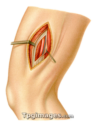
Femoral blood vessel surgery. Historical artwork of the techniques used to expose the femoral (thigh) blood vessels during surgery. This view is of the side of the thigh, with the knee at lower left. The skin (pink) and fat layer (yellow) are shown. Below these, the muscles (dark red) have been dissected, revealing the white membrane surrounding the adductor canal (also called Hunter's canal). This space between the muscles runs down the middle of the thigh. Here, the membrane has been dissected and pulled apart to reveal the femoral artery (red) and vein (blue). Artwork from Atlas and Epitome of Operative Surgery (1898, Otto Zuckerkandl).
| px | px | dpi | = | cm | x | cm | = | MB |
Details
Creative#:
TOP03220563
Source:
達志影像
Authorization Type:
RM
Release Information:
須由TPG 完整授權
Model Release:
N/A
Property Release:
N/A
Right to Privacy:
No
Same folder images:
 Loading