
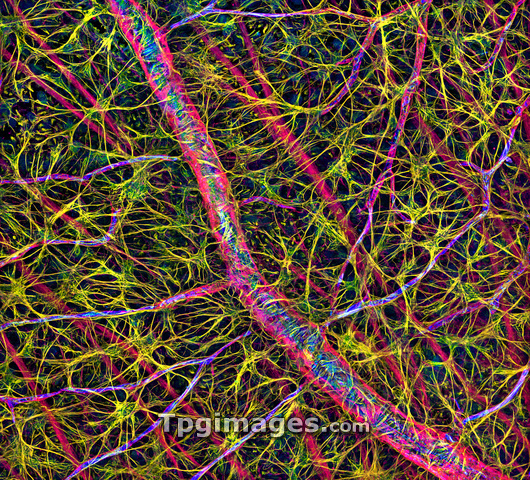
Retina cells. Fluorescent light micrograph of cells in the retina, the light-sensitive membrane that lines the back of the eyeball. A blood vessel runs from top left to bottom right, and numerous other branches are seen. Astrocyte glial cells are yellow. The glial cells provide structural support to the nerve cells that send visual signals to the brain. The tissue has been tagged with fluorescent markers specific to certain proteins. The yellow marks the glial fibrillary acidic protein (GFAP) found in glial cells. Blue marks platelet- endothelial cell adhesion molecule-1 (PECAM-1) found in blood vessels, and red is the structural protein actin.
| px | px | dpi | = | cm | x | cm | = | MB |
Details
Creative#:
TOP03221608
Source:
達志影像
Authorization Type:
RM
Release Information:
須由TPG 完整授權
Model Release:
N/A
Property Release:
N/A
Right to Privacy:
No
Same folder images:
 Loading