
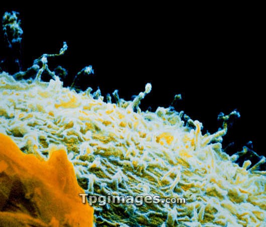
Embryo surface. Coloured scanning electron micrograph of microvilli on the surface of an early embryo. The embryo is at the 2-4 cell stage, the egg has completed only one or two divisions after fertilization. The individual cells in the embryo are called blastomeres and it is a blastomere surface that is seen here. The surface is covered with numerous tiny projections called microvilli. These greatly increase the surface area of the cell and indicate the high metabolic activity of the cell. A probable fragment of a cumulus-corona cell is at bottom left (orange). Magnification: x4,200 at 6x7cm size.
| px | px | dpi | = | cm | x | cm | = | MB |
Details
Creative#:
TOP03222175
Source:
達志影像
Authorization Type:
RM
Release Information:
須由TPG 完整授權
Model Release:
N/A
Property Release:
N/A
Right to Privacy:
No
Same folder images:
 Loading