
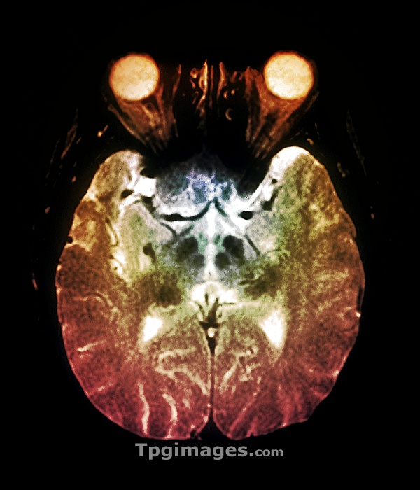
Coloured Magnetic Resonance Imaging (MRI) scan in axial section through the brain of a 65 year old patient, showing Parkinson's disease. At centre are a number of plaques (dark areas) in the thalamus close to the basal ganglia. Parkinson's disease is caused by loss of nerve cells in the basal ganglia resulting in jerky, involuntary movements.
| px | px | dpi | = | cm | x | cm | = | MB |
Details
Creative#:
TOP06659345
Source:
達志影像
Authorization Type:
RM
Release Information:
須由TPG 完整授權
Model Release:
NO
Property Release:
NO
Right to Privacy:
No
Same folder images:
 Loading