
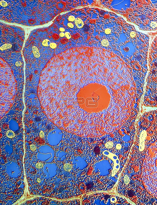
False-colour transmission electron micrograph (TEM) of a cell in the root tip of a maize plant, Zea mays. Plant cells are distinct from animal cells in having an additional external envelope, the plant cell wall, outside the plasma membrane. In this micrograph, the cell wall, made of proteins & polysaccharides, appears as the thin layer between the cells. The wall defines the shape of the cell & expands as it grows. The prom- inent, round organelle in the cell is the nucleus, which contains a smaller, red nucleolus. The yellow areas in the cytoplasm are vacuoles. Magn- ification: x1765 at 35mm size, x2825 at 6x4.5cm size. Reference: MICROCOSMOS, figure 6.1, page 108.
| px | px | dpi | = | cm | x | cm | = | MB |
Details
Creative#:
TOP10166104
Source:
達志影像
Authorization Type:
RM
Release Information:
須由TPG 完整授權
Model Release:
N/A
Property Release:
N/A
Right to Privacy:
No
Same folder images:
 Loading