
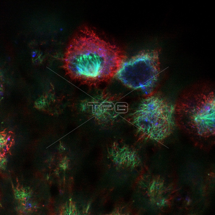
Kidney cells. Confocal fluorescence light micrograph of cultured kidney cells. Antibody- linked dyes have been used to stain tubulin (green), actin (red) and Golgi protein (blue). Tubulin and actin are proteins of the cytoskeleton, the internal framework of a cell involved in cell movement and transport of substances. Golgi proteins are part of the Golgi apparatus, a system of compartments involved in sorting cell products. A confocal microscope only detects light from the focal point of its objective lens. By moving the focal point, images of thin sections of an intact specimen can be obtained. Magnification unknown.
| px | px | dpi | = | cm | x | cm | = | MB |
Details
Creative#:
TOP10188272
Source:
達志影像
Authorization Type:
RM
Release Information:
須由TPG 完整授權
Model Release:
N/A
Property Release:
N/A
Right to Privacy:
No
Same folder images:
 Loading