
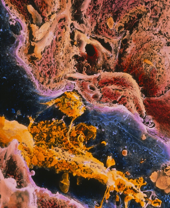
Cirrhosis. Coloured scanning electron micrograph of a human liver cell (red-brown), known as hepatocyte, showing abnormal features caused by cirrhosis. It is a disease in which bands of connective tissue (pale brown upper centre and top left) break up the structure of the liver severely impairing its functions. The surface of the liver cell (upper centre right and left) is abnormal showing irregular microvilli and larger lacunae. Two activated macrophages (Kupffer cells yellow) are seen in a capillary (blue). The wall of the capillary (pale purple) is very thick and fenestrated. Magnification: x1650 at 6x7cm size.
| px | px | dpi | = | cm | x | cm | = | MB |
Details
Creative#:
TOP10196357
Source:
達志影像
Authorization Type:
RM
Release Information:
須由TPG 完整授權
Model Release:
N/A
Property Release:
N/A
Right to Privacy:
No
Same folder images:
 Loading