
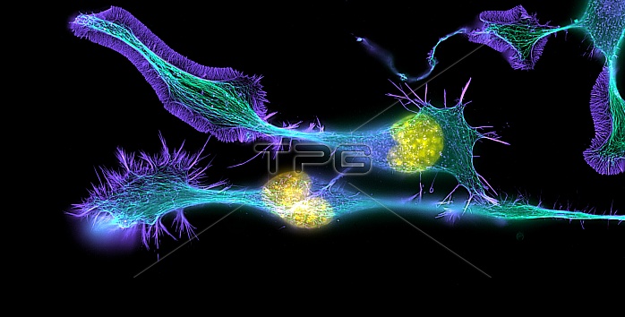
Nerve cancer cells. Immunofluorescence light micrograph of cultured cells from a neuroblastoma, a type of nerve tissue tumour. Fluorescent dyes have been used to highlight proteins in the cell nuclei (yellow) and cytoskeletal protein filaments tubulin (green, forming microtubules) and actin (blue). The cells have been stimulated to undergo neural differentiation, and have changed from a spherical shape to form long branching extensions called neurites (green, blue and purple). These are designed to extend outwards and connect to other neurons. A neuroblastoma is a malignant (cancerous) tumour, most commonly found in children, that derives from primitive nerve cells in a kidney's adrenal gland. Immunofluorescence uses antibodies to attach fluorescent dyes to tissues and molecules in a cell. Magnification: x630 when printed 10cm wide.
| px | px | dpi | = | cm | x | cm | = | MB |
Details
Creative#:
TOP10197496
Source:
達志影像
Authorization Type:
RM
Release Information:
須由TPG 完整授權
Model Release:
N/A
Property Release:
N/A
Right to Privacy:
No
Same folder images:
 Loading