
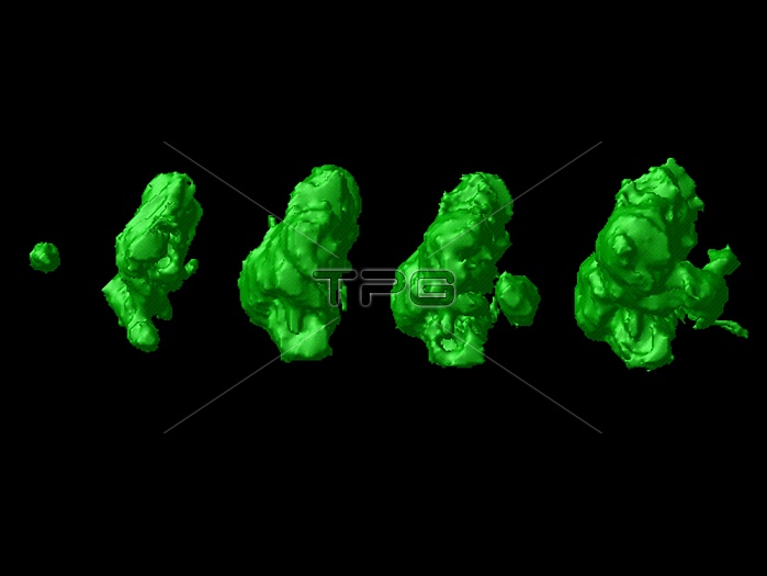
Brain cancer. Coloured three-dimensional (3-D) magnetic resonance imaging (MRI) scans of the growth of a glioblastoma multiforme (GBM) brain tumour. The tumour is seen after diagnosis (day 0, far left), and then at 65, 165, 207 and 249 days. GBM is the most aggressive type of brain cancer. It enlarges rapidly, killing healthy brain cells, and causing a loss of brain function, vomiting, headaches and drowsiness. MRI scans are slice images through the body made using a strong magnet and pulses of radio waves. Successive slices have been combined into a three-dimensional image using a computer. These images were produced at the Laboratory of Neuro Imaging (LONI) at UCLA, USA.
| px | px | dpi | = | cm | x | cm | = | MB |
Details
Creative#:
TOP10197697
Source:
達志影像
Authorization Type:
RM
Release Information:
須由TPG 完整授權
Model Release:
N/A
Property Release:
N/A
Right to Privacy:
No
Same folder images:
 Loading