
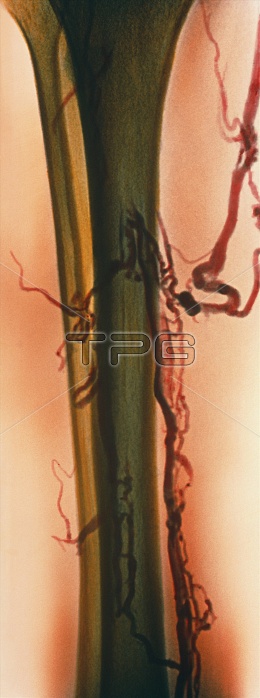
Thrombosis. Coloured venogram (X-ray) showing deep vein thrombosis in the leg's popliteal vein (red, down right). The tibia (shin-bone, olive green) is beside the fibula (centre left). A large thrombus, or abnormal blood clot (dark red, centre right), is in the calf. There are also other smaller clots (dark red) causing irregular blood flow in branching veins of the leg. In thrombosis, a blood clot forms in an undamaged blood vessel, causing pain and tissue damage. Although thrombi often break up on their own, drugs can aid this process and surgery may be needed. A venogram is produced by injecting an X-ray opaque dye into a vein.
| px | px | dpi | = | cm | x | cm | = | MB |
Details
Creative#:
TOP10199347
Source:
達志影像
Authorization Type:
RM
Release Information:
須由TPG 完整授權
Model Release:
N/A
Property Release:
N/A
Right to Privacy:
No
Same folder images:
 Loading