
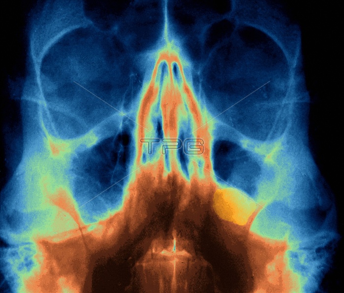
Sinus polyp. Coloured frontal X-ray of the skull of a patient with a polyp growth (yellow, lower right) in the left maxillary sinus. The polyp has developed from the mucous membrane that lines the sinuses. The two maxillary sinuses, like the other paranasal sinuses of the facial bones, are hollow spaces inside bones. The maxillary sinuses are spaces (black, centre left and right) inside the maxilla (upper jaw bones or cheek bones), and lie either side of the nose. The nasal bones (orange, centre) and the eye sockets (black, upper right and left) are also seen. Polyps can turn cancerous and are usually surgically removed.
| px | px | dpi | = | cm | x | cm | = | MB |
Details
Creative#:
TOP10200556
Source:
達志影像
Authorization Type:
RM
Release Information:
須由TPG 完整授權
Model Release:
N/A
Property Release:
N/A
Right to Privacy:
No
Same folder images:
 Loading