
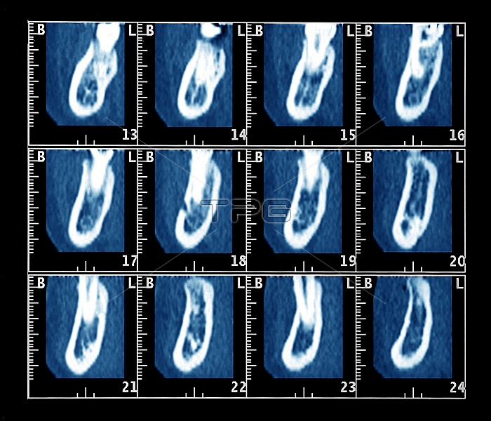
Dental surgery plan. Computed tomography (CT) scans of the lower jaw of a patient, for planning surgery to implant dental prostheses. These are scans 13-24 of 54 sequential vertical scans that show the jaw bone and teeth roots. The vertical plane of the scans is at right-angles to the arc of the jaw, with the front of the teeth at left in each scan. The teeth roots (white, upper centre of most of the scans) are embedded in the jaw bone (white arcs). In this case, the bone appeared too fragile for securing dental implants. See image M780/336 for a horizontal (axial) scan of this jaw that shows the positions of these scans.
| px | px | dpi | = | cm | x | cm | = | MB |
Details
Creative#:
TOP10211863
Source:
達志影像
Authorization Type:
RM
Release Information:
須由TPG 完整授權
Model Release:
N/A
Property Release:
N/A
Right to Privacy:
No
Same folder images:
 Loading