
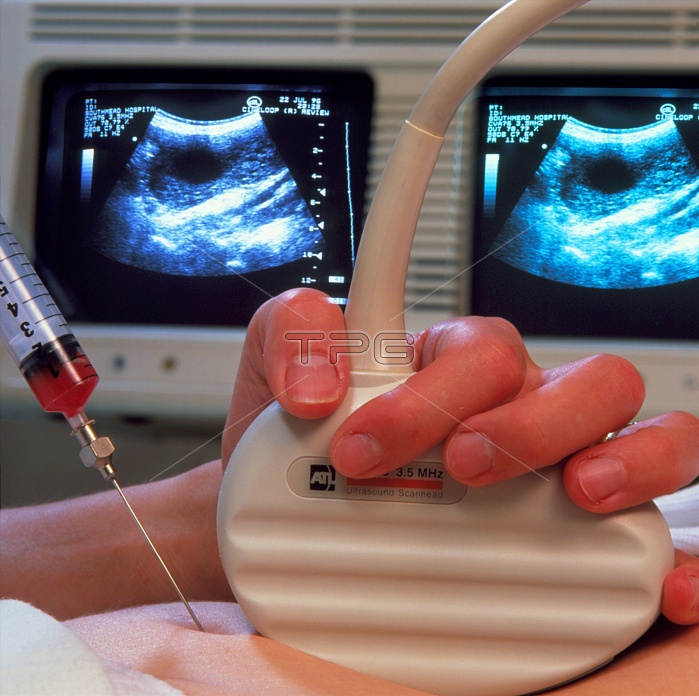
Chorionic villus sampling. Doctor's hand holds an ultrasound emitter (white) onto a woman's pregnant abdomen while drawing a chorionic villus sample into a syringe. On ultrasound screens in the back- ground, the womb of the pregnant woman is seen. The ultrasound imaging enables the needle to be directed to chorionic villi tissue of the foetus which is found in the mother's placenta. Chorionic villi contain foetal blood vessels and is a way of obtaining foetal blood. Chorionic villus sampling (CVS) is usually conducted between 9-12 weeks of pregnancy to evaluate the chromosomal and DNA status of the foetus. CVS is a way of diagnosing abnormalities in a foetus in early pregnancy.
| px | px | dpi | = | cm | x | cm | = | MB |
Details
Creative#:
TOP10212185
Source:
達志影像
Authorization Type:
RM
Release Information:
須由TPG 完整授權
Model Release:
N/A
Property Release:
N/A
Right to Privacy:
No
Same folder images:
 Loading