
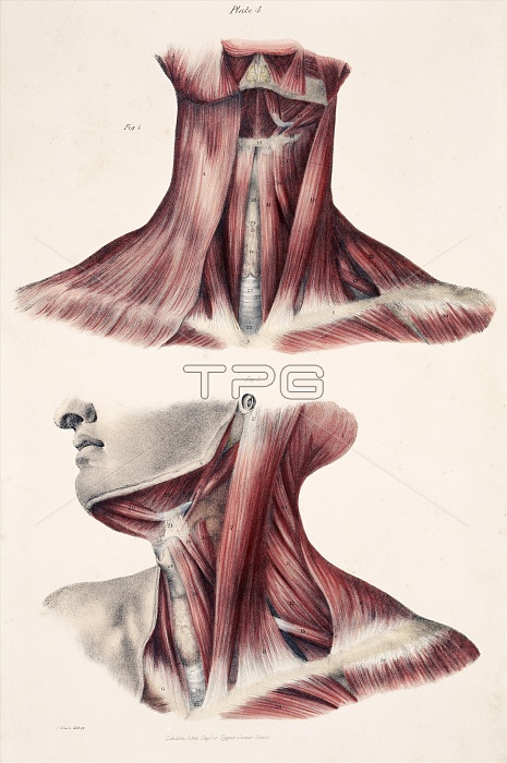
Muscles of the neck, historical artwork. The upper figure shows the front of the neck with the skin and fascia (connective tissue) removed to expose the musculature (red) and tendons. The lower figure shows the muscles on the front and side of the neck as the head is twisted to its right. The left clavicle (collar bone) is exposed on both figures. The trachea (windpipe, white) is in the centre of the neck. Overlying the trachea is the thyroid gland, which secretes hormones to control the rate of metabolism. The major neck muscle from the ear to the base of the neck is the sternocleidomastoid. Published in The Muscles of the Human Body... by Jones Quain in 1836.
| px | px | dpi | = | cm | x | cm | = | MB |
Details
Creative#:
TOP10216202
Source:
達志影像
Authorization Type:
RM
Release Information:
須由TPG 完整授權
Model Release:
N/A
Property Release:
N/A
Right to Privacy:
No
Same folder images:
 Loading