
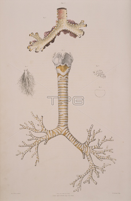
Respiratory tubes. Historical illustration of the respiratory tubes of the lungs. The main diagram at centre shows the trachea (windpipe, yellow and white stripes) dividing into the bronchi of the two lungs. These divide further into bronchioles, eventually forming minute divisions (small diagram at upper left). The end of a bronchiole has been shown magnified (upper right). It has been filled with mercury to show the tiny air sacs (alveoli) where gaseous exchange with the blood takes place. A cross-section of the trachea (centre right) and a rear view of the trachea (top) are also seen. Colour lithograph by Fairland from The Viscera of the Human Body, 1840. Based on drawings by Bagg.
| px | px | dpi | = | cm | x | cm | = | MB |
Details
Creative#:
TOP10216358
Source:
達志影像
Authorization Type:
RM
Release Information:
須由TPG 完整授權
Model Release:
N/A
Property Release:
N/A
Right to Privacy:
No
Same folder images:
 Loading