
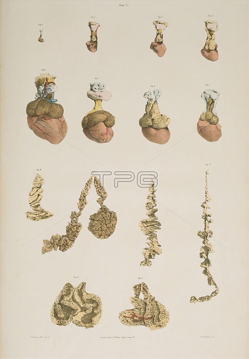
Thymus gland. Historical illustrations of the thymus gland developing from an embryonic state at two months (top left) through successive months to a foetal stage at nine months (upper left). The thymus gland (pink, yellow and brown), the larynx (blue) and the heart (red) are shown. The thymus gland is located at the base of the neck above the heart. It controls the development of lymphoid tissue in infancy and develops the white blood cells of the immune system. The bottom diagrams show the internal and external structure of the thymus gland (normal and unravelled). Colour lithograph by Fairland from The Viscera of the Human Body, 1840. From work by Sir Astley Cooper.
| px | px | dpi | = | cm | x | cm | = | MB |
Details
Creative#:
TOP10216375
Source:
達志影像
Authorization Type:
RM
Release Information:
須由TPG 完整授權
Model Release:
N/A
Property Release:
N/A
Right to Privacy:
No
Same folder images:
 Loading