
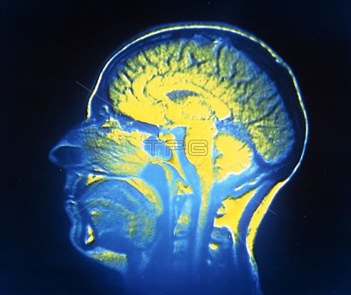
Nuclear Magnetic Resonance (NMR) image of a human head, showing details of the brain, spinal cord, nasal airways & oesophagus. In the brain, the cerebral cortex (the folded outer part) is well- defined; in centre is corpus callosum (which connects the left right cerebral hemispheres). The spinal cord runs down the centre of image; the bulb at its top is the pons, & to its right is the cerebellum (involved in equilibrium & coordination). NMR images are obtained by subjecting the body to a large magnetic field which is interrupted by brief radio pulses, & computing reponse of atoms in different tissues (ionising radiations are not used).
| px | px | dpi | = | cm | x | cm | = | MB |
Details
Creative#:
TOP10219161
Source:
達志影像
Authorization Type:
RM
Release Information:
須由TPG 完整授權
Model Release:
N/A
Property Release:
N/A
Right to Privacy:
No
Same folder images:
 Loading