
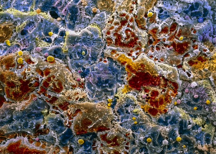
False-colour scanning electron micrograph (SEM) of surface cells on the iris of the eye. The iris is the coloured part of the eye behind the cornea. Pigment cells (melanocytes, blue & brown) can be seen here, joined loosely together by connective tissue fibres (white). Smaller macrophage cells dot the surface. Beneath this matrix (stroma) of iris cells lie muscle fibres. These muscle fibres contract and dilate reflexively, and so control light entering the central pupil of the iris, and falling on the retina. Mobility of the iris is possible through this loose network structure of cells. Magnification: x540 at 6x7cm size. Magnification: x830 at 4x5 inch size.
| px | px | dpi | = | cm | x | cm | = | MB |
Details
Creative#:
TOP10220008
Source:
達志影像
Authorization Type:
RM
Release Information:
須由TPG 完整授權
Model Release:
N/A
Property Release:
N/A
Right to Privacy:
No
Same folder images:
 Loading