
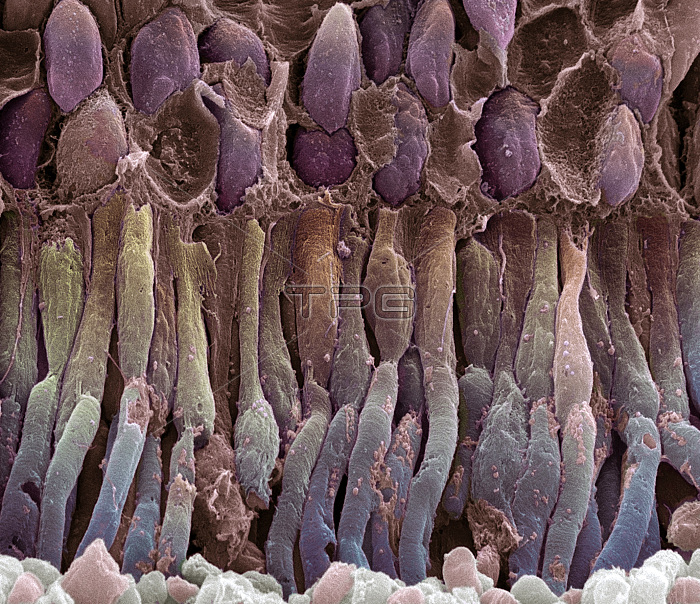
Retina rod cells. Coloured scanning electron micrograph (SEM) of a freeze-fractured section through a retina, revealing the structure of its photoreceptors. Photoreceptors with these rod-like outer parts (blue) are named rod cells (rather than cone cells) and contain the protein rhodopsin (visual purple) that aids vision in dim light. The inner parts (orange) lead to the nuclei (purple, top). Empty spaces in this layer show Muller cells that provide a fibrous support for photoreceptors. Light triggers signals in the photoreceptors after passing through a neuron layer (above nuclei, not seen). These signals travel to the neurons and on to the brain. Magnification: x3000 at 6x7cm size.
| px | px | dpi | = | cm | x | cm | = | MB |
Details
Creative#:
TOP10220056
Source:
達志影像
Authorization Type:
RM
Release Information:
須由TPG 完整授權
Model Release:
N/A
Property Release:
N/A
Right to Privacy:
No
Same folder images:
 Loading