
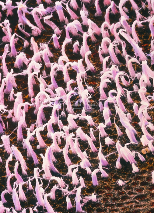
Inner ear. False-colour scanning electron micrograph (SEM) of ciliated hair cells (pink) of the macula utriculi, the neurosensory part of the utriculus, one of the two structures that comprise the membranous labyrinth of the human inner ear. The other structure, the sacculus, is interconnected with the utriculi; both are connected with the subdural space of the brain via a duct, and with the membranous part of the cochlea. The neurosensory areas of both sacculus and utriculi are concerned with the maintenance of equilibrium & with discerning movement and spatial orientation. Magnification: x1150 at 6x7cm size.
| px | px | dpi | = | cm | x | cm | = | MB |
Details
Creative#:
TOP10220172
Source:
達志影像
Authorization Type:
RM
Release Information:
須由TPG 完整授權
Model Release:
N/A
Property Release:
N/A
Right to Privacy:
No
Same folder images:
 Loading