
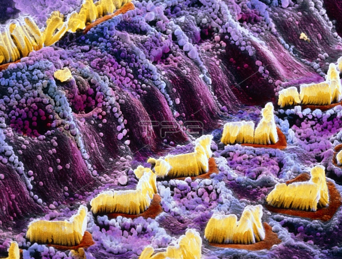
Inner ear. False-colour scanning electron micrograph of hair cells (yellow) which are part of the Organ of Corti in the inner ear. They are divided into outer hair cells (v-shaped) and inner hair cells (top left). Hair cells are immersed in a fluid called endolymph and react to the pressure of sound waves with an undulatory motion. This motion stimulates a nerve ending in each group of hair cells which carries a signal to various parts of the brain via the cochlear nerve. The two types of hair cells are divided by the pillar cells, seen here covered by microvilli (tiny spheres). Magnification: x1320 at 6x7cm size. x2205 at 4x5"~LANDSCAPE"
| px | px | dpi | = | cm | x | cm | = | MB |
Details
Creative#:
TOP10220176
Source:
達志影像
Authorization Type:
RM
Release Information:
須由TPG 完整授權
Model Release:
N/A
Property Release:
N/A
Right to Privacy:
No
Same folder images:
 Loading