
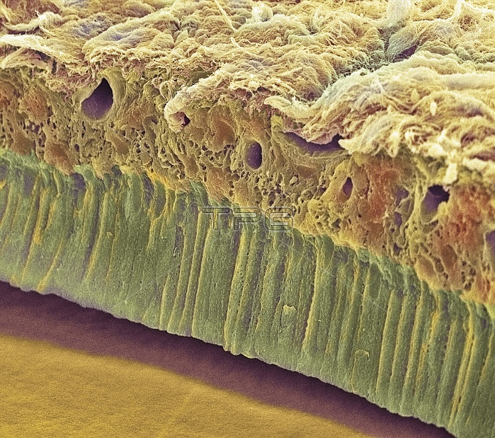
Tooth enamel formation. Coloured scanning electron micrograph (SEM) of a freeze-fractured section through a tooth, showing the enamel-forming cell layer (green). This epithelium comprises a single layer of columnar cells called ameloblasts. The fracture plane passes up into the tooth from the enamel surface (orange, bottom left). The ameloblast layer has detached from the enamel in which it is normally embedded. Enamel is a hard ceramic layer that covers and protects the teeth. The other end of the ameloblasts originate in the internal tooth tissue (brown, across top). Magnification unknown.
| px | px | dpi | = | cm | x | cm | = | MB |
Details
Creative#:
TOP10220397
Source:
達志影像
Authorization Type:
RM
Release Information:
須由TPG 完整授權
Model Release:
N/A
Property Release:
N/A
Right to Privacy:
No
Same folder images:
 Loading