
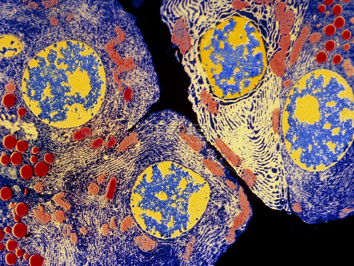
Pancreatic acinar cell. Coloured transmission electron micrograph (TEM) of a slice through several enzyme-secreting acinar cells from a pancreas. The red circles are secretory granules packed with digestive enzymes ready for export to the small intestine. The four round blue and yellow structures are cell nuclei. Each cell is filled with a network of densely folded membrane, called rough endoplasmic reticulum, here coloured light yellow. The membrane's surface is covered in small dots, called ribosomes. These are protein-manufacturing sites where various digestive enzymes are produced and secreted by the cell. Magnification x1200 at 6x4.5cm size.
| px | px | dpi | = | cm | x | cm | = | MB |
Details
Creative#:
TOP10220798
Source:
達志影像
Authorization Type:
RM
Release Information:
須由TPG 完整授權
Model Release:
N/A
Property Release:
N/A
Right to Privacy:
No
Same folder images:
 Loading