
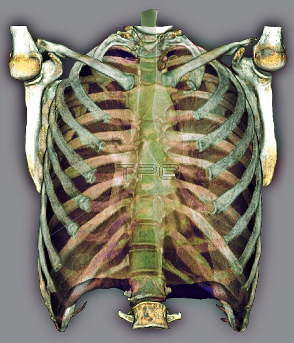
Trachea and rib cage. Coloured frontal 3D computed tomography (CT) scan of a normal trachea and rib cage. The trachea (windpipe, green) runs from the throat (top centre) down to centre, and then splits into the two bronchi, one for each lung (spaces at left and right). The sternum (breast bone) and the rest of the thoracic bones are shown. Twelve pairs of ribs make up the rib cage enclosing the chest. They are attached at one end to the spine (backbone, down centre). The collar bones (clavicles) lie across the top of the rib cage and attach at one end to the breast bone and at the other ends to the shoulder bones. The shoulder blades (scapulas) and the head of the humerus (upper arm bone) are also seen, forming the shoulder joints.
| px | px | dpi | = | cm | x | cm | = | MB |
Details
Creative#:
TOP10221150
Source:
達志影像
Authorization Type:
RM
Release Information:
須由TPG 完整授權
Model Release:
N/A
Property Release:
N/A
Right to Privacy:
No
Same folder images:
 Loading