
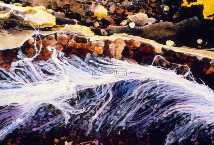
Spermatozoa. False-colour scanning electron micrograph of a bundle of spermatozoa in a seminiferous tubule. The image shows sperm tails with pear-shaped heads visible at bottom. Spermatozoa's formation occurs in the seminiferous tubules in the testes and is divided into two phases called spermatogenesis and spermiogenesis. In the first, the male gamete cells are produced with 23 chromosomes; in spermiogenesis they differentiate into spermatozoa consisting of head, neck and tail. The round brown cells at top are male gametes at the beginning of spermiogenesis. Magnification: x225 at 6x7cm size. x325 at 4x5 inch
| px | px | dpi | = | cm | x | cm | = | MB |
Details
Creative#:
TOP10221283
Source:
達志影像
Authorization Type:
RM
Release Information:
須由TPG 完整授權
Model Release:
N/A
Property Release:
N/A
Right to Privacy:
No
Same folder images:
 Loading