
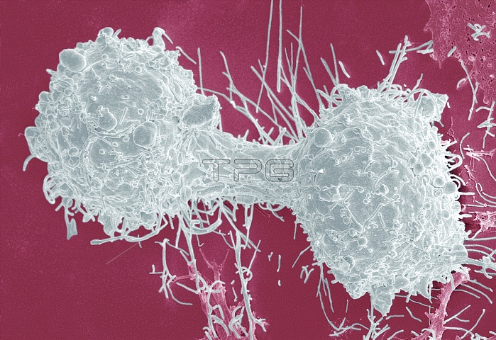
Mitosis. Image 5 of 6. Coloured scanning electron micrograph (SEM) of the surface of a breast cancer cell during cytokinesis, the final stage of mitotic cell division. During mitosis, the nuclear material of a cell divides so that two identical nuclei are formed at opposite ends of a cell. Cytokinesis completes the process by separating the cytoplasm and cell organelles to form two daughter cells (left and right). A ring of actin protein fibres contracts around the centre of the cell, pinching it in two. Magnification: around x2200 when printed 10cm wide. For a sequence of mitosis, see images P672/088 to P672/093.
| px | px | dpi | = | cm | x | cm | = | MB |
Details
Creative#:
TOP10222041
Source:
達志影像
Authorization Type:
RM
Release Information:
須由TPG 完整授權
Model Release:
N/A
Property Release:
N/A
Right to Privacy:
No
Same folder images:
 Loading