
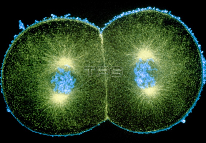
Mitosis. Immunofluorescence micrograph of a newly fertilized sea urchin embryo dividing into two cells. Each of the daughter cells is about to divide again, and is currently in the prophase of mitosis (division of the nucleus). The chromosomes (blue) have condensed and are beginning to align in the middle of a spindle of fibrous microtubules (green) that will pull them apart into two identi- cal sets. The bright green bodies are the poles of the spindles, or centrioles. This picture was made by treating the embryo with fluorescent anti- bodies that bind to certain proteins in the cells. A laser-scanning light microscope was used to cap- ture the image. Magnification unknown.
| px | px | dpi | = | cm | x | cm | = | MB |
Details
Creative#:
TOP10222101
Source:
達志影像
Authorization Type:
RM
Release Information:
須由TPG 完整授權
Model Release:
N/A
Property Release:
N/A
Right to Privacy:
No
Same folder images:
 Loading