
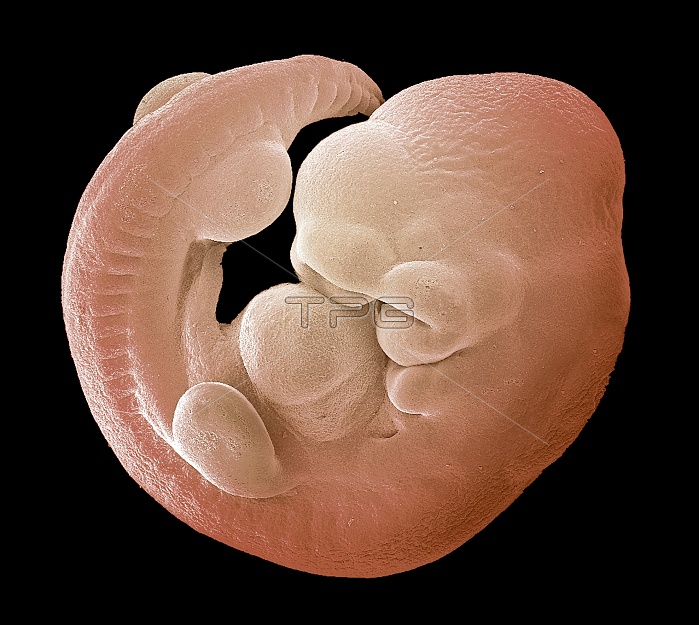
Mouse foetus. Coloured scanning electron micrograph (SEM) of a 17-day-old mouse foetus (Mus musculus). The embryo has started to develop limb buds (left). The head and tail are differentiating and the heart bulge is visible (centre). The embryo attaches to the uterine epithelium about five days after ovulation and the pregnancy lasts for approximately three weeks. Within the uterus the embryo lies inside a fluid-filled sac, known as the amniotic cavity, which is enclosed by double membranes. The fluid protects the embryo against the pressure of the internal organs of the mother and from any injury from the mother's movements.
| px | px | dpi | = | cm | x | cm | = | MB |
Details
Creative#:
TOP10222554
Source:
達志影像
Authorization Type:
RM
Release Information:
須由TPG 完整授權
Model Release:
N/A
Property Release:
N/A
Right to Privacy:
No
Same folder images:
 Loading