
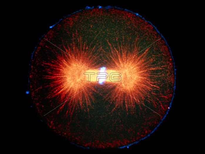
Mitosis. Immunofluorescence micrograph of a newly fertilized sea urchin embryo during the metaphase of mitosis (division of the nucleus). This cell is about to divide into two. The chromosomes (blue) have aligned in a band across the middle of a "spindle" of fibres called microtubules (orange). These will pull the chromosomes apart into two identical sets to form the nuclei of the daughter cells. The bright orange bodies at left and right are the poles of the spindle, or centrioles. This picture was made by treating the cell with fluor- escent antibodies that bind to specific proteins. The image was then captured by a laser-scanning light microscope. Magnification unknown.
| px | px | dpi | = | cm | x | cm | = | MB |
Details
Creative#:
TOP10277750
Source:
達志影像
Authorization Type:
RM
Release Information:
須由TPG 完整授權
Model Release:
NO
Property Release:
NO
Right to Privacy:
No
Same folder images:
 Loading