
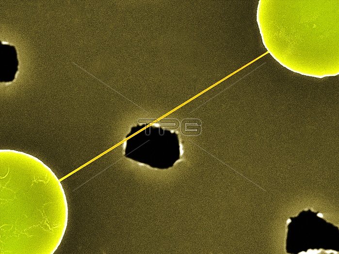
DNA bundle on silicon nanopillars. Coloured scanning electron micrograph (SEM) of a DNA (deoxyribonucleic acid) bundle and silicon nanopillars used to obtain the first high-contrast direct images of DNA. The DNA bundle was prepared on a bed of silicon nanopillars (bottom left and top right). The holes allow the passage of electrons for transmission electron microscopy (TEM). The bundle (several DNA strands) is around 20 nanometres wide. This image was obtained in 2012 by a team led by Enzo di Fabrizio from the Department of Nanostructure, at the Italian Institute of Technology (IIT) in Genoa. For the TEM image, see C015/3216.
| px | px | dpi | = | cm | x | cm | = | MB |
Details
Creative#:
TOP10403265
Source:
達志影像
Authorization Type:
RM
Release Information:
須由TPG 完整授權
Model Release:
No
Property Release:
No
Right to Privacy:
No
Same folder images:
 Loading