
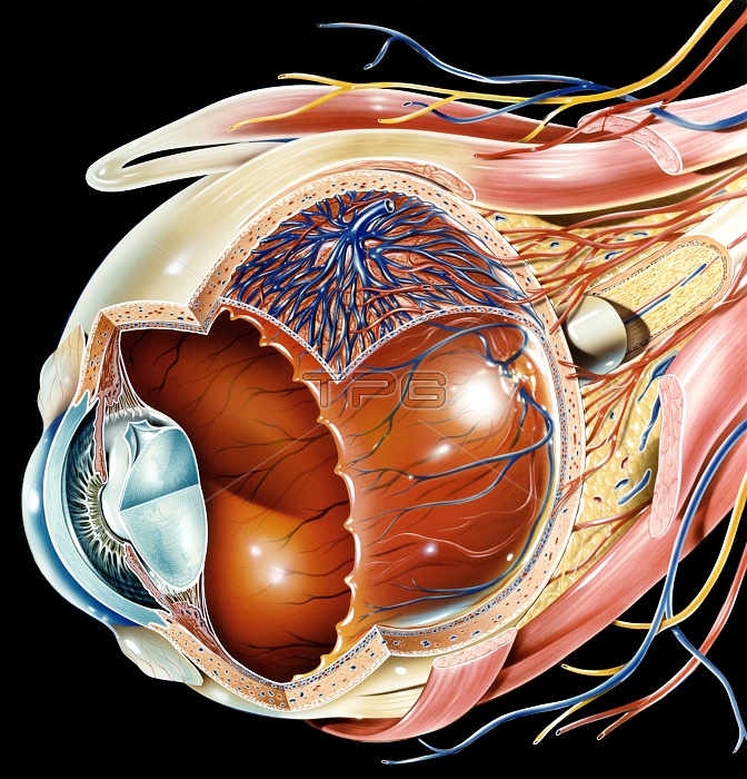
Eye anatomy, artwork. The eye is seen in cutaway format from the side. Structures shown include the lens (light blue, lower left), its attachment points, the iris, and the bulge of the cornea at the front of the eye. Several layers of the eyeball are shown, including the sclera (outer layer), the choroid, and the retina (innermost). Retinal capillaries (red and blue) are shown. The optic nerve (yellow) is at far right. External structures shown include muscles (red) that control the movement of the eyeball, and nerves (yellow) and more arteries (red) and veins (blue).
| px | px | dpi | = | cm | x | cm | = | MB |
Details
Creative#:
TOP11718508
Source:
達志影像
Authorization Type:
RM
Release Information:
須由TPG 完整授權
Model Release:
NO
Property Release:
NO
Right to Privacy:
No
Same folder images:
EYEORGANTISSUEHUMANBODYANATOMYBIOLOGYOPHTHALMOLOGYNEUROLOGYARTWORKILLUSTRATIONCUTAWAYBLACKBACKGROUNDNORMALHEALTHYOPHTHALMICOCULAREYEBALLDISSECTEDDISSECTIONVISIONVISUALSENSESENSORYSIGHTLENSIRISCORNEACORNEALSCLERAWHITEOFTHEEYECHOROIDRETINARETINALBLOODVESSELSVEINSARTERIESCAPILLARIESCAPILLARYBEDVENOUSARTERIALVASCULAROPTICNERVEMUSCLESNERVESARTERYVEINANATOMICALBIOLOGICALNEUROLOGICAL
ANATOMICALANATOMYARTERIALARTERIESARTERYARTWORKBACKGROUNDBEDBIOLOGICALBIOLOGYBLACKBLOODBODYCAPILLARIESCAPILLARYCHOROIDCORNEACORNEALCUTAWAYDISSECTEDDISSECTIONEYEEYEEYEBALLHEALTHYHUMANILLUSTRATIONIRISLENSMUSCLESNERVENERVESNEUROLOGICALNEUROLOGYNORMALOCULAROFOPHTHALMICOPHTHALMOLOGYOPTICORGANRETINARETINALSCLERASENSESENSORYSIGHTTHETISSUEVASCULARVEINVEINSVENOUSVESSELSVISIONVISUALWHITE
 Loading