
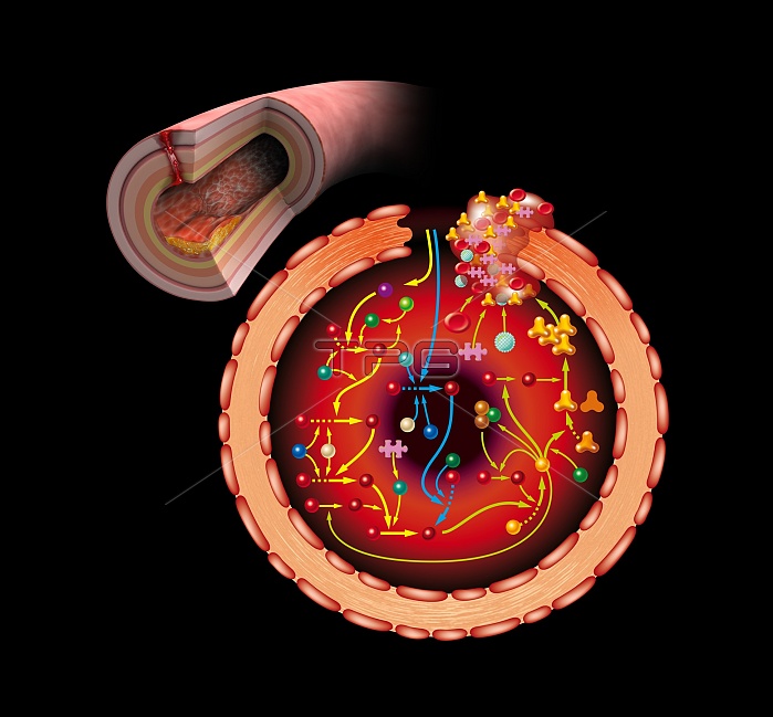
Blood coagulation cascade. Artwork of the biochemical cascade of blood chemicals and proteins during blood clotting (coagulation). The blood vessel and its layered wall is at upper left. The main diagram shows a clot forming at the damaged area (upper right). This occurs via reaction chains known as pathways mediated by factor X proteins (red spheres). The extrinsic pathway (down centre, blue arrows) and intrinsic pathway (down left, yellow arrows) unite in the common pathway (up right, from thrombin, yellow sphere) to form the blood clot. This consists of fibrin (yellow trefoils), platelets (pink puzzle pieces), red blood cells (red discs), and white blood cells (light blue spheres).
| px | px | dpi | = | cm | x | cm | = | MB |
Details
Creative#:
TOP11719635
Source:
達志影像
Authorization Type:
RM
Release Information:
須由TPG 完整授權
Model Release:
NO
Property Release:
NO
Right to Privacy:
No
Same folder images:
 Loading