
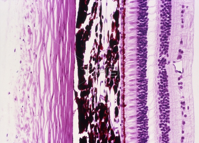
Eye wall. Light micrograph of a cross section through the eye wall. The outer layer (left, pink) is the sclera, connective tissue arranged in bundles. The next layer (centre, dark red) is the choroid. This layer contains blood vessels and a pigment to absorb excess light to prevent blurred vision. Inside of the choroid is the multi-layered retina. Its first layer is a line of pigment cells (red), immediately followed by the light sensitive rod and cone cells (pink). For light to reach these cells it must pass through layers of nerve cells. The nerve cell nuclei are visible as layers of purple dots. Haematoxylin and eosin stained. Magnification x100 at 35mm size.
| px | px | dpi | = | cm | x | cm | = | MB |
Details
Creative#:
TOP11722263
Source:
達志影像
Authorization Type:
RM
Release Information:
須由TPG 完整授權
Model Release:
NO
Property Release:
NO
Right to Privacy:
No
Same folder images:
 Loading