
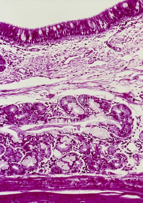
Windpipe. Light micrograph of a section through the windpipe (trachea). The windpipe is a tube taking air to the lungs. At top (white) is the lumen. Lining the windpipe (top, dark pink) is the epithelium containing goblet cells producing mucus. The epithelial cells are covered with tiny hair-like cilia (just visible) which with mucus remove dust from the air. The next layer (spongy appearance) is the lamina propria, connective tissue with a rich blood supply. The submucosa below is also connective tissue and it contains numerous glands (pink, rounded). At bottom (dark pink) is the surface of the underlying cartilage. Haematoxylin and eosin stained.
| px | px | dpi | = | cm | x | cm | = | MB |
Details
Creative#:
TOP11722283
Source:
達志影像
Authorization Type:
RM
Release Information:
須由TPG 完整授權
Model Release:
NO
Property Release:
NO
Right to Privacy:
No
Same folder images:
 Loading