
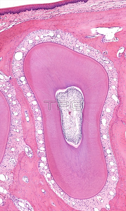
Light microscopy of a tooth. This section is below the gumline and shows how the root of the tooth is bordered by a connective tissue zone called the periodontal ligament. It suspends the tooth in a socket of jaw bone stained pink. Spaces in the ligament are blood vessels and nerves are also present. The centre of the tooth is the pulp cavity (root canal) that also has vessels and nerves. Most of the tooth seen here is dentin, a hard tissue consisting of fine, long tubules that contain collagen, proteins and mineralized matrix. Magnification x75 when printed at 10 cm.
| px | px | dpi | = | cm | x | cm | = | MB |
Details
Creative#:
TOP15070432
Source:
達志影像
Authorization Type:
RM
Release Information:
須由TPG 完整授權
Model Release:
No
Property Release:
No
Right to Privacy:
No
Same folder images:
 Loading