
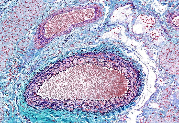
Light microscopy of two muscular arteries. This is the most common type among arteries. Starting innermost is the internal elastic layer/lamina (part of the tunica intima) showing folding due to vessel contraction when fixed for histology. Next is multi-layered smooth muscle (tunica media) and an outer thinner connective tissue layer with some elastic fibres called the tunica adventitia. Muscular arteries serve to distribute and maintain blood flow to organs and tissues using their smooth muscle to control vessel diameter. Magnification x140 when narrow width printed at 10 cm.
| px | px | dpi | = | cm | x | cm | = | MB |
Details
Creative#:
TOP15205072
Source:
達志影像
Authorization Type:
RM
Release Information:
須由TPG 完整授權
Model Release:
No
Property Release:
No
Right to Privacy:
No
Same folder images:
 Loading