
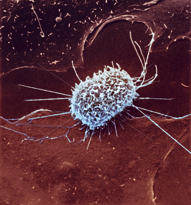
Dividing cancer cell. Coloured scanning electron micrograph (SEM) of a cultured cancerous (malignant) cell from ovary tissue, showing the microvilli (finger-like projections) covering its surface. This surface appearance is typical, but not definitive, for cultured cells during the anaphase stage of mitotic cell division.
| px | px | dpi | = | cm | x | cm | = | MB |
Details
Creative#:
TOP15339877
Source:
達志影像
Authorization Type:
RM
Release Information:
須由TPG 完整授權
Model Release:
N/A
Property Release:
N/A
Right to Privacy:
No
Same folder images:
1abnormalanaphasecancercancerouscellcelldivisionchinesehamstercoloredcolouredconditionculturecultureddiseasediseaseddisorderdividingfalse-coloredfalse-colouredhamstertissuehealthcarehistopathologicalhistopathologymalignantmammalmammalianmedicalmedicinemicrovillimicrovillusmitosismitoticoncologicaloncologyoneovarianovaryscanningelectronmicrographscanningelectronmicroscopesemsinglesurfacestructureunhealthy
1abnormalanaphasecancercancerouscellcellchinesecoloredcolouredconditionculturecultureddiseasediseaseddisorderdividingdivisionelectronelectronfalse-coloredfalse-colouredhamsterhamsterhealthcarehistopathologicalhistopathologymalignantmammalmammalianmedicalmedicinemicrographmicroscopemicrovillimicrovillusmitosismitoticoncologicaloncologyoneovarianovaryscanningscanningsemsinglestructuresurfacetissueunhealthy
 Loading