
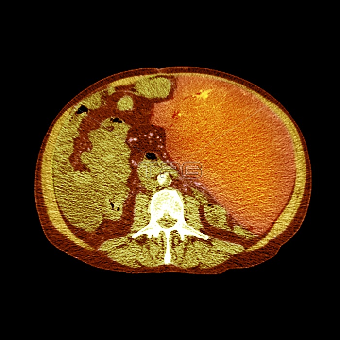
Secondary peritoneal cancer. Coloured computed tomography (CT) scan of an axial section through the abdomen and pelvis of a 71-year-old male patient with a recurrent gastrointestinal stromal tumor (GIST), showing numerous large peritoneal deposits throughout the abdomen and peritoneal cavity, and multiple cancerous lesions that have spread (metastasised) to the liver. GISTs are rare tumours that develop in mesenchymal tissue (muscle, connective tissues and bone) with symptoms that include trouble swallowing (dysphagia), gastrointestinal haemorrhage (bleeding) or secondary tumours. The peritoneum is the serous membrane that forms the lining of the abdominal cavity. Also present here is moderate volume ascites (accumulation of fluid in the peritoneal cavity).
| px | px | dpi | = | cm | x | cm | = | MB |
Details
Creative#:
TOP15941051
Source:
達志影像
Authorization Type:
RM
Release Information:
須由TPG 完整授權
Model Release:
N/A
Property Release:
N/A
Right to Privacy:
No
Same folder images:
 Loading