
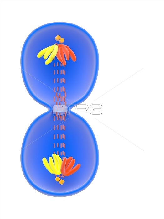
Cytokinesis in animal cells. Illustration of cytokinesis, the final stage of cell division, in an animal cell. The cell's genetic material (DNA) in the form of chromosomes (red and yellow) has separated into chromatids that have moved to opposite ends of the dividing cell. They are near centrioles (orange), to which they are attached by short kinetochore fibres (red dashed lines). Similar microtubules (red, down centre) condense to form a rod called the midbody. At centre, a contractile ring of actin and myosin is dividing the cell into two. In addition, the nuclear envelope is reforming around the chromosomes. For this artwork with labels, see C023/8854.
| px | px | dpi | = | cm | x | cm | = | MB |
Details
Creative#:
TOP15984392
Source:
達志影像
Authorization Type:
RM
Release Information:
須由TPG 完整授權
Model Release:
N/A
Property Release:
N/A
Right to Privacy:
No
Same folder images:
 Loading