
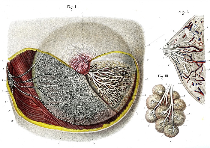
Breast anatomy. 1866 illustrations showing the anatomy of a human breast. Fig. 1: cutaway of the breast, showing the lymph vessels (white lines, left), milk (lactiferous) ducts (white, branching, right), nipple (centre) and surrounding pectoral muscle (brown). Fig. 2: section through the breast, showing the milk ducts (white lines) leading to the nipple (left), with the underlying mammary tissue. Fig. 3: A lobule of alveoli (round) from the mammary tissue, with milk ducts (white).
| px | px | dpi | = | cm | x | cm | = | MB |
Details
Creative#:
TOP16029547
Source:
達志影像
Authorization Type:
RM
Release Information:
須由TPG 完整授權
Model Release:
N/A
Property Release:
N/A
Right to Privacy:
No
Same folder images:
1800s186619thcenturyalveolialveolusanatomicalanatomyareolaartworkbiologicalbiologybreastbreastscirculatorysystemcutoutcutoutscut-outcut-outscutoutcutoutsductsfemaleglandshistoricalhistoryhistoryofsciencehumanbodyillustrationlactiferousductslobulelymphvessellymphaticsystemmammaryglandmilkductnippleno-onenobodyorganorganspectoralmusclereproductivesystemskinstructurestructuressystemtissuevesselswhitebackgroundwoman
1800s186619thalveolialveolusanatomicalanatomyareolaartworkbackgroundbiologicalbiologybodybreastbreastscenturycirculatorycutcutcut-outcut-outscutoutcutoutsductductsductsfemaleglandglandshistoricalhistoryhistoryhumanillustrationlactiferouslobulelymphlymphaticmammarymilkmusclenippleno-onenobodyoforganorgansoutoutspectoralreproductivescienceskinstructurestructuressystemsystemsystemsystemtissuevesselvesselswhitewoman
 Loading