
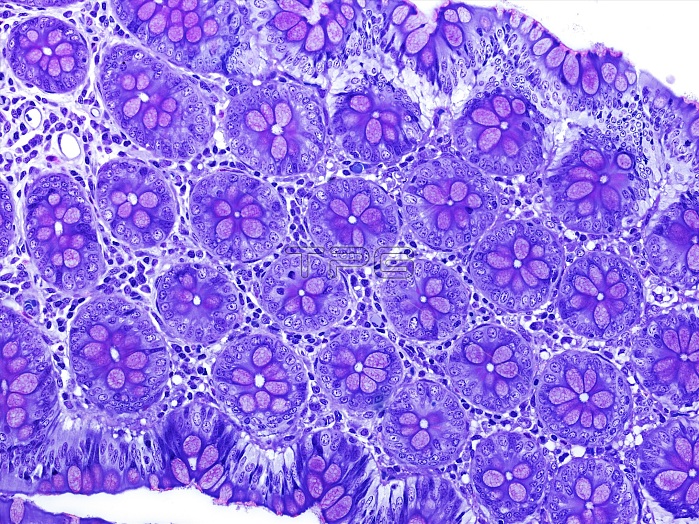
Small intestine tissue. Light micrograph of a longitudinal section through tissue from the small intestine. This view shows cross-sections through many intestinal glands called crypts of Lieberkuhn. Crypts are long blind-ending tube-like extensions of the surface epithelial lining of the gut. In the small intestine they comprise several cell types including mucus-secreting goblet cells (purple) and absorptive enterocytes (blue) around a narrow central lumen. Crypts also contain gut epithelial stem cells. Connective tissue supporting the crypts contains fibroblasts, nerves, blood vessels, and white blood cells. Magnification: x183 when printed at 10 centimetres across.
| px | px | dpi | = | cm | x | cm | = | MB |
Details
Creative#:
TOP16633402
Source:
達志影像
Authorization Type:
RM
Release Information:
須由TPG 完整授權
Model Release:
N/A
Property Release:
N/A
Right to Privacy:
No
Same folder images:
 Loading