
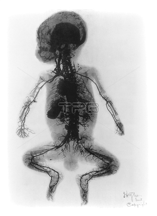
X-ray arteriogram of a child (1899). Early X-ray showing in fine detail the arteries in a child's body. The X-ray was made in 1899 at St. Thomas' Hospital in London, and published in the Archives of the Roentgen Ray. It was titled: "An injected infant". The cadaver of a young boy was injected, at the femoral artery of the leg, with four pounds of mercury. Under X-ray, arteries of the limbs, abdomen, heart, neck and face are highlighted. This important diagnostic technique was later to become known as an arteriogram or angiogram. It took, however, many years before harmless X-ray opaque substances were developed which could be injected to show arteries in living patients.
| px | px | dpi | = | cm | x | cm | = | MB |
Details
Creative#:
TOP19257216
Source:
達志影像
Authorization Type:
RM
Release Information:
須由TPG 完整授權
Model Release:
N/A
Property Release:
N/A
Right to Privacy:
No
Same folder images:
 Loading