
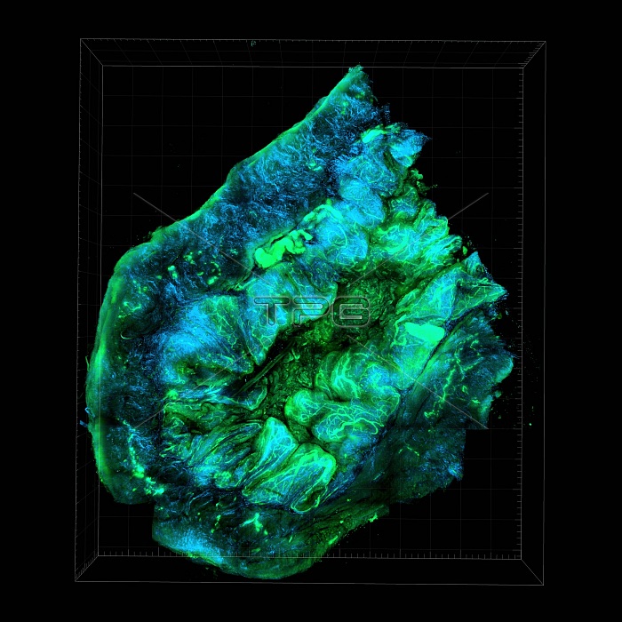
Ovarian cancer. Optical tissue clearing image of tissue from an ovarian cancer, showing the interplay of collagen and blood vessels. This is done using second harmonic signals (blue) and autofluorescent signals (green; green fluorescent protein, GFP). This is a murine SKOV cell line tumour seeded with CD63+ cells, revealing the tumour-stromal interfaces that comprises the tumour microenvironment (TME). The image demonstrates the interplay of collagen II fibrils and blood vessels generated from angiogenesis. The tumour microenvironment is being studued because it plays a crucial role in helping cancers to grow and evade destruction. This image is of a mouse model of ovarian cancer.
| px | px | dpi | = | cm | x | cm | = | MB |
Details
Creative#:
TOP19460865
Source:
達志影像
Authorization Type:
RM
Release Information:
須由TPG 完整授權
Model Release:
N/A
Property Release:
N/A
Right to Privacy:
No
Same folder images:
 Loading