
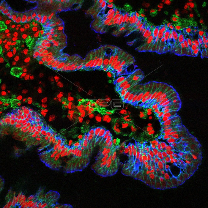
Small intestine villus. Fluorescence light micrograph of a cross-section through a villus from a normal human small intestine mucosa. The intestinal villi are finger-like projections which increase the surface area of the intestine, improving the efficiency of food absorption. Integrin, which can be found on the basolateral membrane of epithelial cells, and also shows up on scattered stromal cells, is stained in green. Cytokeratin, which stains epithelial cells, is shown in blue, and cell nuclei are stained in red.
| px | px | dpi | = | cm | x | cm | = | MB |
Details
Creative#:
TOP20028515
Source:
達志影像
Authorization Type:
RM
Release Information:
須由TPG 完整授權
Model Release:
N/A
Property Release:
N/A
Right to Privacy:
No
Same folder images:
NO-ONENOBODYFLUORESCENTFLUORESCINGCELLULARCELLSNORMALHEALTHYBIOLOGICALHISTOLOGICALCROSS-SECTIONSECTIONEDCELLNUCLEICYTOSKELETONCYTOSKELETALINTESTINESDIGESTIVESYSTEMTRACTGASTROINTESTINALEPITHELIALMUCOSAVILLICRYPTSOFLIEBERKUHNCRYPTOFLIEBERKUHNINTEGRINBASOLATERALMEMBRANESTROMALCELLSCYTOKERATINEPITHELIALCELLSPROTEINPROTEINSSINGLETISSUECELLVILLUSINTESTINESMALLINTESTINEHUMANBODYCELLBIOLOGYBIOLOGYGASTROENTEROLOGYHISTOLOGYFLUORESCENCELIGHTMICROGRAPHLMLIGHTMICROSCOPE1ONE
1BASOLATERALBIOLOGICALBIOLOGYBIOLOGYBODYCELLCELLCELLCELLSCELLSCELLSCELLULARCROSS-SECTIONCRYPTCRYPTSCYTOKERATINCYTOSKELETALCYTOSKELETONDIGESTIVEEPITHELIALEPITHELIALFLUORESCENCEFLUORESCENTFLUORESCINGGASTROENTEROLOGYGASTROINTESTINALHEALTHYHISTOLOGICALHISTOLOGYHUMANINTEGRININTESTINEINTESTINESLIEBERKUHNLIEBERKUHNLIGHTLIGHTLMMEMBRANEMICROGRAPHMICROSCOPEMUCOSANO-ONENOBODYNORMALNUCLEIOFOFONEPROTEINPROTEINSSECTIONEDSINGLESMALLSTROMALSYSTEMTISSUETRACTVILLIVILLUSINTESTINE
 Loading