
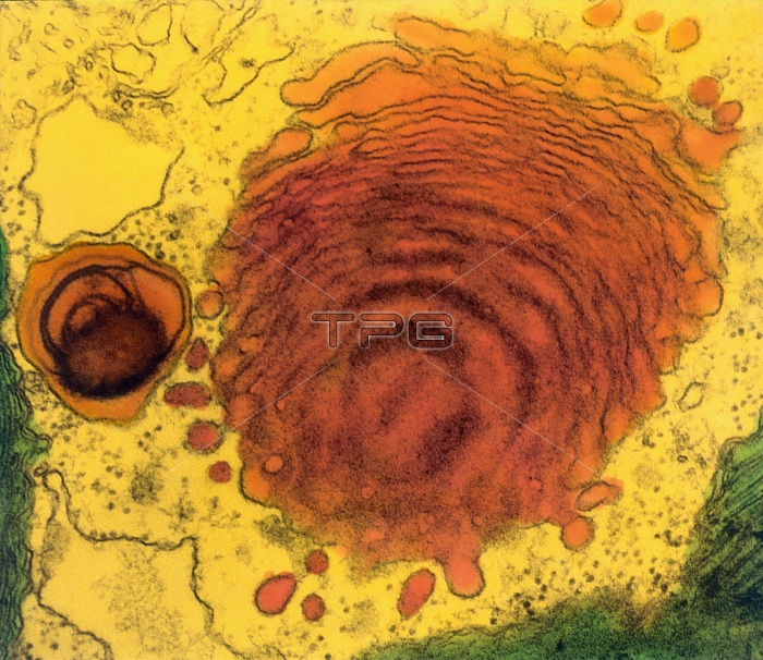
Color-enhanced Transmission Electron Micrograph (TEM) showing lysosome and Golgi body. A small single-membraned lysosome with lipid contents (pink) lies beside a larger Golgi body (also pink) in Euglena; vesicles are budding from the edges of the Golgi cisternae into the cytoplasmic matrix (yellow); lysosomes are also formed from the Golgi complex and are filled with hydrolytic enzymes used to digest macromolecules during metabolism and aging. Magnification: 30,000X at 2.25" film size.
| px | px | dpi | = | cm | x | cm | = | MB |
Details
Creative#:
TOP22214038
Source:
達志影像
Authorization Type:
RM
Release Information:
須由TPG 完整授權
Model Release:
N/A
Property Release:
No
Right to Privacy:
No
Same folder images:
 Loading