
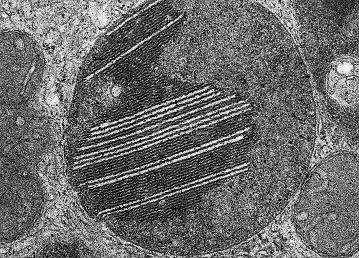
Transmission electron micrograph of lysosomes in the interstitial cells of Leydig in the testis of the domestic boar. Additional examples of laminated inclusions in lysosomes are illustrated here in micrographs from Leydig cells which, like other steroid-secreting cells, are rich in lysosomes. The clear spaces interposed among the lamellae are negative images of thin tabular crystals of unknown nature that have been dissolved in the course of specimen preparation.
| px | px | dpi | = | cm | x | cm | = | MB |
Details
Creative#:
TOP22218878
Source:
達志影像
Authorization Type:
RM
Release Information:
須由TPG 完整授權
Model Release:
N/A
Property Release:
No
Right to Privacy:
No
Same folder images:
 Loading