
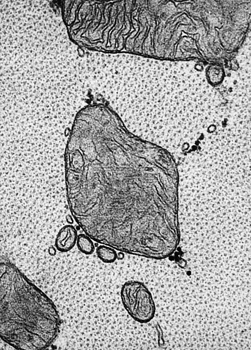
Transmission electron micrograph of papillary muscle from the heart. This transverse section passes through the region of filaments interdigitating at the end of the A band. One can identify the myosin filaments on end as a uniformly distributed population of large dots, each surrounded by a number of smaller dots representing the cut ends of the actin filaments.
| px | px | dpi | = | cm | x | cm | = | MB |
Details
Creative#:
TOP22219266
Source:
達志影像
Authorization Type:
RM
Release Information:
須由TPG 完整授權
Model Release:
N/A
Property Release:
No
Right to Privacy:
No
Same folder images:
 Loading