
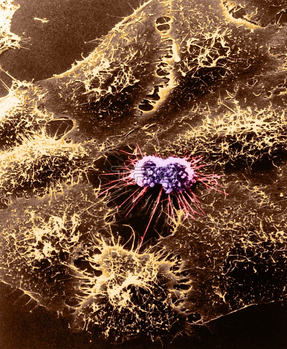
Color enhanced scanning electron micrograph of cultured HeLa cells originally derived many years ago from a woman's cancerous cervical tissue. This HeLa cell has been infected with adenovirus. After 4-1/2 hours the HeLa cell's surface becomes rough. The multiple surface blebs on this cell characteristic for a certain stage of cell division that both normal and cancer cells go through. Research with the SEM has established the extraordinarily responsive nature of cell surface form. This instrument records, in pictures, specific cell reactions to various changes in the cells environment and maps the distribution of surface binding sites for biologically important molecules such as hormone, antigens, and pharmacologic agents.
| px | px | dpi | = | cm | x | cm | = | MB |
Details
Creative#:
TOP22223294
Source:
達志影像
Authorization Type:
RM
Release Information:
須由TPG 完整授權
Model Release:
N/A
Property Release:
No
Right to Privacy:
No
Same folder images:
 Loading